
Does preservation make sense before we know how to revive?

My name is Aurelia Song and I hope to make whole-body, human, end-of-life preservation for future revival a new global tradition. I care about it so much I’ve dedicated my life to it.1
The biggest objection I get to end-of-life preservation goes like this: “We can’t revive today, so we can’t prove that preservation works. Therefore preservation probably doesn’t work. We shouldn’t bother with preservation until we can revive.” I call this the immediate revival objection.
I respect the immediate revival objection. If your standard of evidence is full recovery, then you don’t need any knowledge of how people or mental processes work on the inside to evaluate preservation; you can just observe that they survive a round trip.
I think requiring revival, now, is reasonable a priori—it’s analogous to how I feel when people talk about new kinds of quantum computers: I’ll believe it when they’re actually doing something useful.
However, in my opinion the logic of the immediate revival objection is too conservative when it comes to end-of-life preservation. Instead, I think that as a scientific community, we’ve known enough to preserve people for at least 30 years. I think we can and should start preserving people today. I think if you knew what I know, you’d agree.
Why do I think that? What’s my response to the immediate revival objection? How do I know what I think I know?
The San Diego Frozen Zoo
Dr. Kurt Benirschke started the San Diego Frozen Zoo in 1975. It just celebrated its 50th anniversary. Benirschke had the visionary idea to preserve cells from endangered animal species using liquid nitrogen, with the belief that people in the future would probably want them. DNA had been shown to be a durable, double-helix-shaped polymer in 1953, and successful cryopreservation of sperm had been achieved in 1949, so Dr. Benirschke was in a position to understand that animal cells each separately held the “molecule of heredity” and were preservable by cold.
That’s all he needed to begin preserving. And today, after 50 years in storage, samples are now starting to be used.
Think of the state of our biological knowledge when the San Diego Frozen Zoo opened: no one understood what DNA meant at a programmatic level, no one had cloned a mammal, and Kary Mullis wouldn’t go on his fateful LSD trip and invent PCR for another decade. The idea of using preserved DNA to revive an extinct species, in 1975, probably sounded just as far-fetched as scanning and simulating a preserved brain does today.2
Dr. Benirschke preserved cells anyway. He didn’t need to know exactly how they would be used. He did his job well and gave us, today, an option we wouldn’t otherwise have.
What can we learn from the example of the San Diego Frozen Zoo? There’s two lessons I take from it:
Only basic knowledge is needed to preserve. You might think that you need to have masterful knowledge of the thing you’re preserving in order to preserve it. But Dr. Benirschke didn’t have masterful knowledge of DNA, he only had basic knowledge, and that was clearly enough. The amount of knowledge needed to preserve something is often vastly less than that needed to actually do anything with that you’re preserving.
Preservation can work before you can revive. You might think that if you don’t know everything about what you’re preserving, then you need to at least be able to “unpreserve” to have anything worthwhile. But the San Diego Frozen Zoo preserved animal cells before the invention of PCR, before the first successful cloning of a mammal, before they had conclusive proof that what they were preserving would be useful. I’m sure they faced criticism along the lines of “how could a few cells ever be useful for species conservation?”, “how could we ever fit a whole genome on a computer?”, “why are you wasting money on something that might never be used?”. These questions turned out to be focused on the wrong thing. What mattered was whether preservation captured the necessary information. The founders of the Frozen Zoo didn’t know exactly how their cells would be used, and they didn’t need to in order for their project of preservation to be useful. In the words of Dr. Kurt Benirschke, “you must collect things for reasons you don’t yet understand.”
The time to start preserving is when you’re reasonably confident you can do the preservation, not after you’ve demonstrated how to use what you’re preserving.3 The San Diego Frozen Zoo successfully preserved animal cells in the early 70s, before anyone knew very much about what DNA meant or how proteins folded or how to manipulate DNA. They didn’t know how or if the cells they were preserving would ever be used. But despite their ignorance, the chemical and biological knowledge of the 1970s was up to the modest task of showing that preserving even a few animal cells almost certainly preserved many copies of the animal’s genome, and that a few preserved cells would likely be sufficient to “remember” what a species was, and that was clearly the right time to start.4
I believe that today when it comes to preserving people we’re in an analogous position with neuroscience as we were with genetics in the 1970s: We have more than sufficient knowledge to confidently preserve, but not enough to do much with what we’re preserving. Yet.
Preserving People
Why do I think the lessons of the San Diego Frozen Zoo apply to human end-of-life preservation?
I’ll start with a neuroscience overview. What’s your brain physically made of? How does it store information? I’ll only cover the basics, the stuff that was already well-established more than 20 years ago, because we don’t need more than the basics. Like Dr. Benirschke of the San Diego Frozen Zoo, we don’t need a complete understanding of neuroscience to know enough to preserve.
Then I’ll talk about how fixation physically works and what it can and can’t preserve.
Next I’ll briefly touch on Deep Hypothermic Circulatory Arrest, which I’ve written about before. DHCA demonstrates that we don’t have to preserve dynamic brain activity, only structure, because people recover from having that activity “zeroed out”.
Finally I’ll put it all together in information-theoretic terms and develop a formal definition of adequacy for a preservation technique. Fixation’s good at preserving structure, but is it good enough? We’ll use the framework to evaluate whether fixation can preserve what we really care about: people.
What does neuroscience say about how the brain encodes information structurally?
Top level summary: it turns out that to create a long-term behavioral change, neuroscience says you must physically change multiple synapses. Synapses, broadly, are the durable physical trace of memory we’re looking to preserve.
Important: I don’t need neuroscience to be “complete” to evaluate whether preservation works. I need to understand the basics of what the brain’s made of and what’s physically different between different people. These are basic facts we need to know, and we’ve had the basics for a while—none of it has changed for decades. This section is here, not to review advanced neuroscience, but to celebrate that we really do know the basics of the brain enough to justify preservation.
Conversations with neuroscientists
If you walk up to a neuroscientist and say: “hey we know a lot about neuroscience, so how soon before we upload the first person like I saw in ‘Pantheon‘?”, that neuroscientist will probably say something along the lines of “We know nothing about the brain. We’re so far from uploading that it won’t happen for 100 years. There are major open questions in neuroscience about how the brain works, individual cells have vast complexity, and we can’t even simulate a C. elegans yet. No one knows how memory works—we’re still working on decoding even the simplest memories and there are all kinds of theories.”
Now imagine you walk up to that same neuroscientist and say: “No one knows anything about the brain! Despite the efforts of science it remains a complete mystery! For all we know, a rock could be conscious. Maybe even the whole universe is conscious! Isn’t that neat?” That neuroscientist would probably say something like: “What do you mean ‘we don’t know anything about the brain’? We know a lot about the brain! Neuroscientists have done 75 years of amazing work since Hodgkin and Huxley figured out how the ionic dynamics of the action potential worked. We can erase memories by altering synapses. We can create false memories in mice using optogenetics. We’ve spent decades working out how the biochemistry of synapses works and we have what amounts to a ‘parts list’ at a proteomic level. We’ve mapped the fruit fly connectome and accurately simulated its visual system. We actually know quite a bit about how the brain works, how memories are formed, how it processes information. There are a lot of mysteries, sure. We don’t know how a lot of stuff works at a systems level. But we know a lot about the basics. A rock is not conscious.”
What’s inside your head?
Let’s talk about what scientists do know about the brain, starting with its basic anatomy. When you open up a skull and look inside, what do you see?
The large-scale: white matter and grey matter
First off, your brain has two obviously different parts to it: 500 ml of white matter and 650 ml of grey matter. There’s also around 200 ml of apparently empty space (ventricles) filled with clear fluid (cerebral spinal fluid, or CSF) (Irimia 2021).
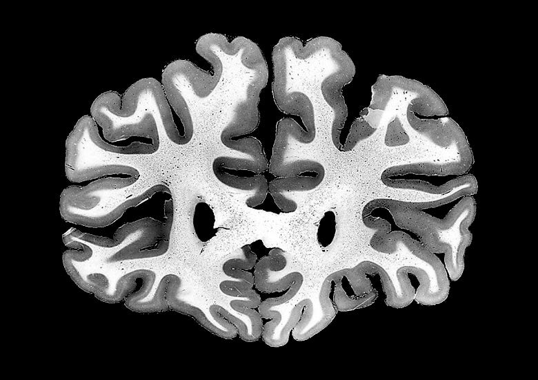
The white matter, visually, looks like a vast bundle of wires connecting the grey matter to other parts of itself. The grey matter is where the neuron cell bodies live. You may think, as I used to, that neurons are very small. This is not the case! A single projection neuron in the right hemisphere might grow an axon that extends across the corpus callosum and connects with another neuron in the grey matter of the left hemisphere. That’s a single cell that’s 10 cm long! The bundles of fibers in the white matter are all literally extensions of the neurons in the grey matter.
There’s some blood in your brain in addition to the white and grey matter, but probably not as much as you think. All the blood vessels in the brain amount to about 50 ml in total.5 Around half is capillaries, each itself as wide as a single red blood cell, and the other half is larger blood vessels. Only capillaries are thin enough to allow the interchange of oxygen and sugar between blood and brain which nourishes each of your neurons.6 Capillaries penetrate every part of the brain, and a brain cell is never very far from one. Capillaries are much denser in the grey matter (~5.5% of volume) where the cell bodies are, and sparser in the white matter (~1.5% of volume) (Lu 2005, Gould 2016).
What does the brain look like at a microscopic level?
The vast majority of the grey matter, around 75%, is “neuropil”, with less than 15-25% being cell bodies and blood vessels (Cano-Astorga 2024) .
That’s a lot of volume dedicated to “neuropil”! What’s neuropil made of? It’s almost entirely synapses, axons, dendrites, and glial cells. Synapses, axons, and dendrites are all different parts of the anatomy of the neurons, whose cell bodies live in the grey matter. Axons are, broadly, the “output” part of the neuron, and dendrites are the “input” part. Synapses are what join the outputs to the inputs.
Within the neuropil itself, axons and dendrites each take up ~33% each of the total volume (Karbowski 2015). Glial processes take up ~14% of the volume. Synapses take up the remaining ~20% (Wilhelm 2014).7
White matter has far fewer cell bodies and blood vessels compared to grey matter, being almost entirely composed of long-range “wires” (axons) connecting neurons in different regions of grey matter across centimeters. (Coelho 2018). It’s essentially neuropil that’s all “output”.
What about energy use?
Energy is not used frivolously in biology. If something is using energy, it’s because it’s doing something important. Doubly so if it’s using a disproportionate amount of energy.
How does a person spend their internal energy? First off, the brain is hungry! Your brain consumes 20% of your body’s energy, but it’s only around 2% of your body’s mass (Mink 1981). The brain is important energetically.
How does the brain spend its energy? At a high level, the white matter takes about 25% and the grey matter takes the other 75%, despite them being approximately the same volume (Harris 2012). The grey matter is important energetically.
How does the grey matter spend its energy? First, around 25% of the total energy is used to continuously rebuild the proteins and other macromolecules of each cell (housekeeping). About 15% of the energy is used to create action potentials, and an additional 15% is used to maintain baseline polarization of neurons at around -70 mV (keeping the lights on). The rest of the energy (45%) is used to power synapses (Howarth 2012), despite them being 20% of the grey matter’s volume. Like the brain itself, synapses consume disproportionately large amounts of energy.
Synapses seem important!
Synapses are each around half a femtoliter in volume (Wilhelm 2014, Benavides-Piccione 2012) and you have around 250 trillion in total (Tang 2001).8
What do those hundreds of trillions of synapses, using so much of the brain’s energy, do?
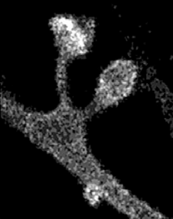
Synapses change when memories change
Synapses change shape when memories are formed (Choi 2021). The physical changes synapses undergo in response to learning aren’t subtle, often doubling or halving their size (Matsuzaki 2004). You can label which synapses change during memory formation, and if you “reset” just those synapses, you erase that specific memory, and not other memories learned both shortly before and after the memory you delete (Hayashi-Takagi 2015). When you disrupt synaptic plasticity machinery, you can temporarily prevent long-term memory formation (Goto 2021). You can create false memories by artificially strengthening new synapses (Vetere 2019). Synapses operate at millisecond timescales and nerve impulses travel quickly throughout the brain and body, which are exactly the right dynamics for the speed of our thoughts. Synapses can last a lifetime (Bhatt 2009, Yang 2009).
Here’s what synapses look like at a molecular level
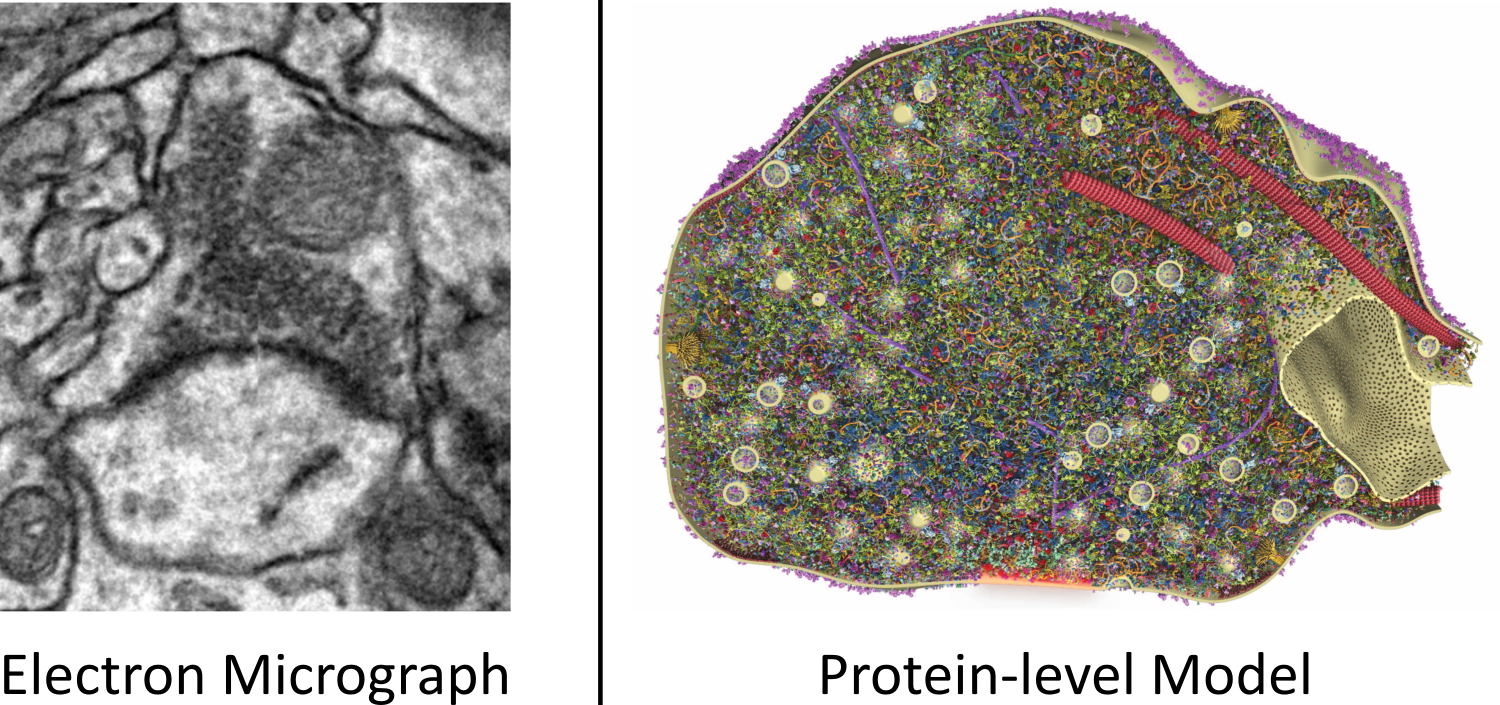
I didn’t appreciate this when I first started preserving brains, but a synapse (as well as every cell in the body) is absolutely full of proteins, as you can see in the picture above (which again is only showing around 1/3rd of the proteins!). Before I saw models like this, I thought that cells were mostly empty bags of water with proteins elegantly doing their thing, gliding past each other with plenty of “elbow room” between proteins, as is often depicted in many visualizations. I now find myself surprised that cells are even liquid at all, instead of solid peptide blocks.9
Synapses changes size by fractions of a femtoliter in response to memory formation, which changes their “strength” (how much they influence the neuron to which they connect, electrophysiologically). What changes when a synapse changes size by a fraction of a femtoliter? A volume like that is small at our scale, but huge at an atomic scale. If a synapse expands by half a femtoliter, it’s adding roughly 500,000 additional proteins, each containing around 10,000 individual atoms (Wilhelm 2014).10
Synapses are durable
We saw in my previous post that the surgical procedure called deep hypothermic circulatory arrest (DHCA) cools people to 16°C, stops respiration and circulation, effectively zeros out the dynamic electrical state of the nervous system, yet doesn’t erase long-term memory. We need some durable physical substrate of memory that survives cooling to explain this. Do synapses physically survive the cooling used during DHCA, unlike the brain’s electrical activity? Yes! (Xie 2012)
Synapses are the physical basis of learning and memory
When we look inside the brain we see, essentially, two-hundred-and-fifty trillion femtoliter-sized switches which grow and shrink by fractions of a femtoliter in response to learning.
The actual cell bodies in the grey matter don’t change in response to learning. Neither do the long-range connections in the white matter, or the blood vessels spread throughout the brain. But in the neuropil, we see that it’s synapses that change in response to learning, while being physically robust enough to survive DHCA.11 That’s why I believe that synapses are the physical trace of memory. What does the broader neuroscience literature say?
What do neuroscience review papers and textbooks say?
The field of neuroscience is broadly in consensus, and has been for many decades at this point: synapses are the physical basis of learning and memory.
I think it’s worth understanding just how established this picture of the mind and brain is. So here are twenty distinct sources from noteworthy neuroscience papers and textbooks that all point to the same bottom line. (Emphasis added):
“Perhaps the most striking finding in the cell biology of memory is that the consolidation and long-term storage of memory involves transcription in the nucleus and structural changes at the synapse. These structural components of learning-related synaptic plasticity can be grouped into two general categories: (1) remodeling and enlargement of preexisting synapses, and (2) alterations in the number of synapses, including both the addition and elimination of synaptic connections.”
Bailey, Kandel, and Harris 2015“The classic view is that items are embedded in long term memory via specific synaptic modifications, and presentation of these items leads to activation of stable activity patterns in the network (‘attractors’).”
Barak and Tsodyks 2014“Learning is primarily mediated by activity-dependent modifications of synaptic strength within neuronal circuits.”
Bittner et al. 2017“[The] ability of synapses to individually change their structure and composition in a long-lasting way is an essential mechanism for synaptic plasticity and represents the cellular basis of learning and memory.”
Bosch et al. 2014“Today, it is generally accepted that the neurobiological substrate of memories resides in activity driven modifications of synaptic strength and structural remodeling of neural networks activated during learning.”
Bruel-Jungerman et al. 2007“Long-lasting changes in the synaptic connectivity between neurons are generally accepted to be crucial for the establishment and maintenance of memories.”
Gobbo et al. 2017“It is generally believed that changes in the synaptic connections between neurons play a major role in learning and memory formation. While short-term memory might rely mainly on the strengthening and weakening of pre-existing synapses, long-term storage of information is thought to require structural reorganization of neuronal networks, the formation of new synapses and the loss of existing connections.”
Hofer and Bonhoeffer 2010“One of the chief ideas we shall develop in this book is that the specificity of the synaptic connections established during development underlie perception, action, emotion, and learning.”
Kandel et al. Principles of Neural Science 2021“Synaptic plasticity is generally accepted as the principal implementation of information storage in neural systems.”
Kukushkin and Carew 2017“In the quest for the physical substrate of learning and memory, a consensus gradually emerges that memory traces are stored in specific neuronal populations and the synaptic circuits that connect them.”
Lu & Zuo 2021“From a neural circuit point of view, learning is a process to transform a neural network to adapt to the environment, and memory is the state of maintaining such a network… Various forms of synaptic plasticity, the persistent change in synaptic efficacy, are widely believed to be the cellular substrate underlying learning and memory. Among them, long-term potentiation (LTP) and long-term depression (LTD), two opposite forms of synaptic plasticity, have been studied most extensively. LTP and LTD were initially discovered by electrophysiological recording, but subsequent research has revealed accompanying morphological changes in dendritic spines.”
Ma & Zuo 2021“It is widely believed that encoding and storing memories in the brain requires changes in the number, structure, or function of synapses… This axiomatic view that synaptic plasticity is critical for learning and memory is supported by data derived from many different memory systems, neural circuits, and molecular pathways mediating an array of different behaviors.”
Maren 2005“Changing the strength of connections between neurons is widely assumed to be the mechanism by which memory traces are encoded and stored in the central nervous system… We conclude that a wealth of data supports the notion that synaptic plasticity is necessary for learning and memory…”
Martin, Grimwood, and Morris 2000“We now understand in considerable molecular detail the mechanisms underlying long-term synaptic plasticity and the importance that such plastic changes play in memory storage, across a broad range of species and forms of memory. One surprising finding is the remarkable degree of conservation of memory mechanisms in different brain regions within a species and across species widely separated by evolution.”
Mayford, Siegelbaum, and Kandel 2012“Memories are believed to be stored as long-lasting structural changes in synapses.”
Moczulska et al. 2013“[I]n the last 10 years findings from this field have provided key contributions towards establishing the idea that stable, long-lasting changes in synaptic function underlie learning and memory.”
Silva 2003“Considerable evidence suggests that the formation of long-term memories requires a critical period of new protein synthesis… Studies in mammals have demonstrated that bidirectional changes in synaptic growth accompany synaptic plasticity.
Sutton & Schuman 2006“At the molecular level, the formation and consolidation of long-term memory are thought to be ultimately expressed in the form of structural changes at synapses.”
Wittenberg, Sullivan & Tsien 2002“Our findings reveal that rapid, but long-lasting, synaptic reorganization is closely associated with motor learning. The data also suggest that stabilized neuronal connections are the foundation of durable motor memory.”
Xu et al. 2009“The obvious site to compactly store information is at the synapse. Storage occurs by changing its transfer ‘weight,’ that is, its ability to excite or inhibit a postsynaptic neuron. Since the synapse is the key site for processing information, storing it there avoids additional wire for relay. Moreover, information stored directly at a synapse can be retrieved directly—also avoiding additional wire. In short, as we peruse a blueprint of brain design, we should not seek a special organ for ‘information storage’—it is stored, as it should be, in every circuit.” (Chapter 14)
Sterling and Laughlin 2017 Principles of Neural Design
What does chemical fixation do?
Our protocol for preserving people at Nectome calls for chemical fixation of every cell via vascular perfusion of aldehydes. We use an aldehyde called glutaraldehyde to achieve fixation. It’s fixation that’s our primary and most important method for achieving preservation.
I believe that fixation, as used in Nectome’s method, preserves the microscopic and large-scale anatomy of a person’s brain and body, including, importantly, all synapses. I believe that in addition to structure, fixation additionally preserves almost all biological macromolecules present in a person’s entire body including proteins, nucleic acids like DNA and RNA, and lipids, in almost the same configuration they had during life.
Why do I believe this?
What does glutaraldehyde actually do?
Glutaraldehyde is a kind of aldehyde (formaldehyde is also an aldehyde) used to preserve tissue. You couldn’t see a single molecule of glutaraldehyde if you tried to accurately draw it in any of the previous images in this post, because it would take up less than 1% of a pixel even in the earlier image showing synaptic proteins. Glutaraldehyde has a molecular weight of 100.12 g/mol, while the individual proteins pictured are around 30,000 g/mol. During preservation, we flood the vascular system with glutaraldehyde. It crosses cell membranes in seconds (Leung 2001, Walter 1986) and starts crosslinking proteins to themselves and to other proteins. After around 60 seconds, the cytoplasm forms a gel that traps essentially all proteins, DNA, lipids, etc in-place (Huebinger 2018).
Why do I believe that proteins are preserved?
Immunohistochemistry
Why do I think that proteins are still present after fixation? Mainly because we can still observe proteins after fixation: the field of immunohistochemistry is built on measuring the positions and amounts of proteins in cells using antibody staining. One of the first steps of preparation of tissue for immunohistochemistry is to fix proteins with aldehydes. Check out the Supplemental Figures from (Wilhelm 2014), the same paper the protein-level synapse model from the last section is from. Those researchers studied the vesicle transport proteins that make up about 1/3rd of the total proteins by weight in a synapse. That’s ~300,000 total proteins split into 62 different kinds of proteins. (A synapse has around 1,000-2,000 different kinds of proteins and ~1,000,000 total proteins in half a femtoliter.) For each of those 62 proteins, they used antibodies after fixation to find where they are inside the synapse. That shows that the proteins are still there and that they’re still identifiable with antibody labeling. Not a single one of the proteins they examined was removed by the fixation process, and those proteins were selected based on being part of vesicle transport, not being able to be preserved by glutaraldehyde, so it’s likely most other proteins are likewise preserved.
Bulk protein measurements
What if, in a hypothetical world, fixation just removed half of all the proteins but kept the other half? Then immunohistochemistry might find that “all the different kinds of proteins are present” even though a substantial number are lost in an absolute sense. How can we distinguish between “extractive fixation” and “comprehensive fixation”?
I believe that most of the “stuff” present before preservation is still there after fixation, because bulk protein measurements can’t measure a difference in protein content between fixed vs frozen tissue. In (Ostasiewicz 2010), the researchers took rats and measured the protein content of fresh-frozen brain tissue vs the protein content of brain tissue that they fixed and then paraffin embedded, which includes total removal of all water and many lipids via alcohol and xylene dehydration and infiltration of paraffin wax into the tissue. Here’s their results:
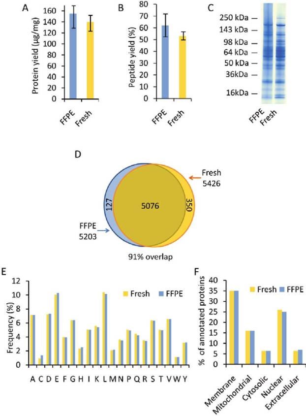
Protein content after fixation is my second-favorite null result in science.12 We’ll get to my favorite later.
Other biological macromolecules like lipids (Morgan 1967, Leist 1986) and DNA (Tokuda 1990)13 are also retained during fixation.
Why do I believe microscopic anatomy is preserved?
It’s useful to know that biomolecules are likely preserved by fixation, but it’s possible that biomolecules would be retained while the microscopic anatomy is scrambled. How do we know, for example, whether synapses are created, destroyed, or moved during fixation, even while the underlying biomolecules are preserved? Suppose that during the 60 seconds of fixation before the cytoplasm gels and further microscopic movement becomes impossible, that a neuron writhes and randomly disconnects and reconnects its synapses? That might would result in some potentially normal-looking microanatomy and all proteins retained, but in reality the preserved microstructure would not accurately reflect the living microstructure. How do we know, when we look at seemingly well-preserved tissue, that the connections we see are the same connections that were present before preservation?
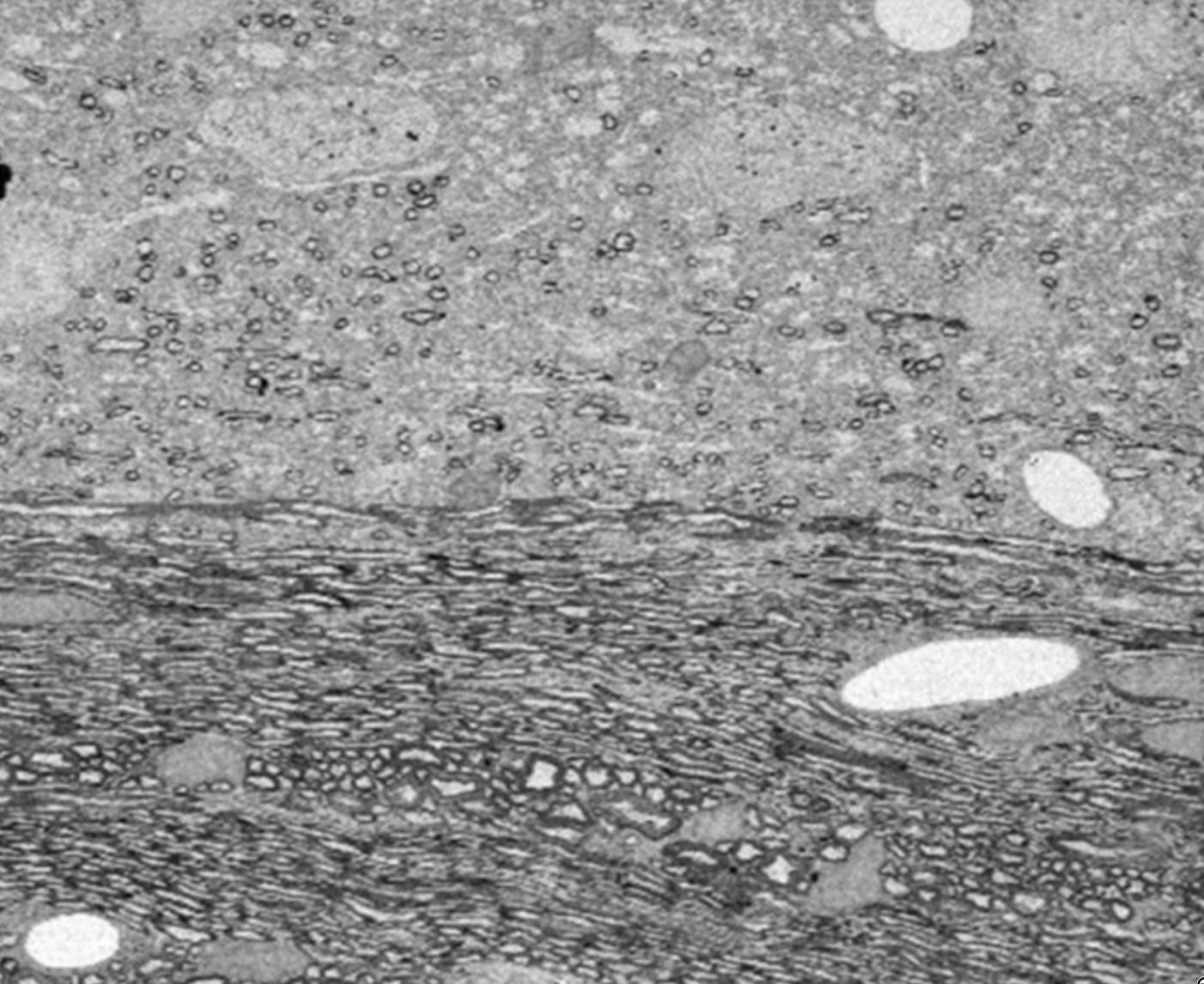
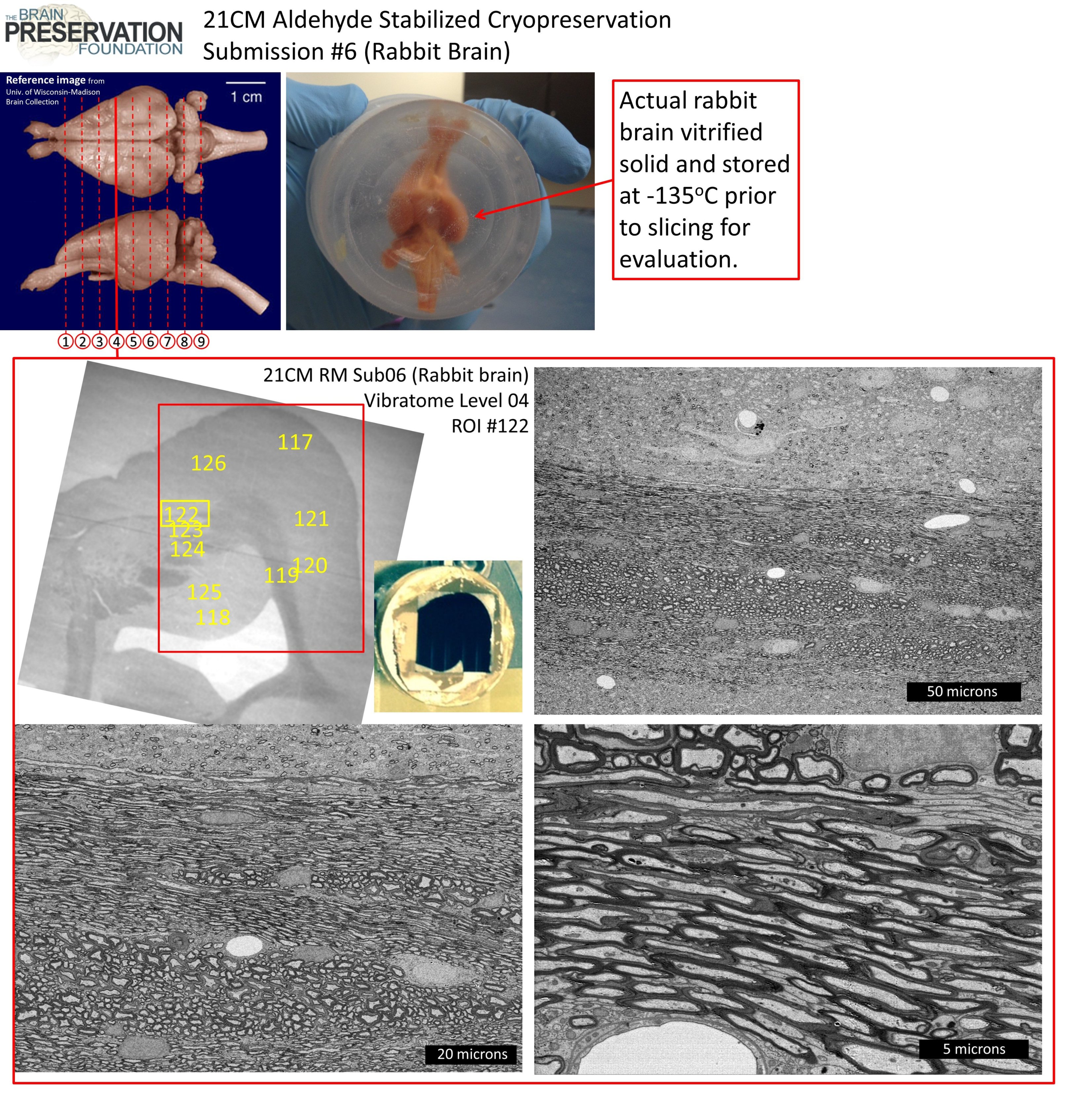
I believe that fixation preserves the brain’s microanatomy because of the correlative microscopy studies that have been done where researchers used superresolution two-photon microscopy to take a picture of a section of a single neuron and its synapses, preserved the entire brain with fixatives, and then found that exact neuron again and imaged it with electron microscopy.
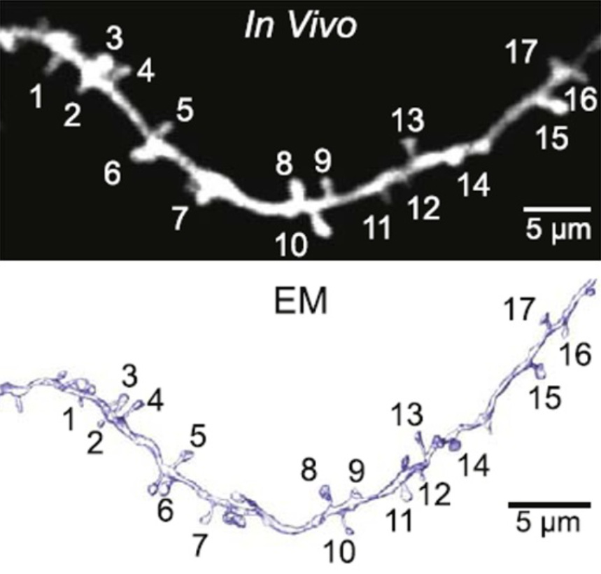
This isn’t a one-off image, it’s an example taken from a paper from the large and growing field of Correlated Light and Electron Microscopy (CLEM). This particular paper impressed the Brain Preservation Foundation enough to be nominated for its Aspirational Neuroscience Prize.
I’d be surprised if fixation altered the brain’s synaptic connections—I don’t know any compelling first-principles reason for fixation to disrupt synapses during crosslinking (on the contrary, it should stabilize them), and when people directly measure physical changes from fixation, they find the same synapses before and after. Fixation directly altering synapses in a way that still looks anatomically normal afterward would, however, be the kind of thing that could invalidate Nectome’s preservation protocol, even in spite of us winning the brain preservation prize, so I take it seriously.
Deep Hypothermic Circulatory Arrest teaches us that we don’t need to preserve dynamic activity
So far we’ve talked about the physical structure of the brain and fixation’s ability to preserve that structure. But what about the dynamics—the second-to-second changes in ion concentration, neurotransmitters, voltages, etc?
I don’t believe dynamic activity is necessary to preserve. I realize this is an extremely convenient belief for someone who runs a preservation company that’s really good at preserving structure and unable to preserve dynamics. But the causality actually goes the other way: when I first learned that dynamic brain activity can be zeroed out without loss of information, in Sebastian Seung’s intro to neuroscience class at MIT, that’s what actually got me interested in preservation in the first place.
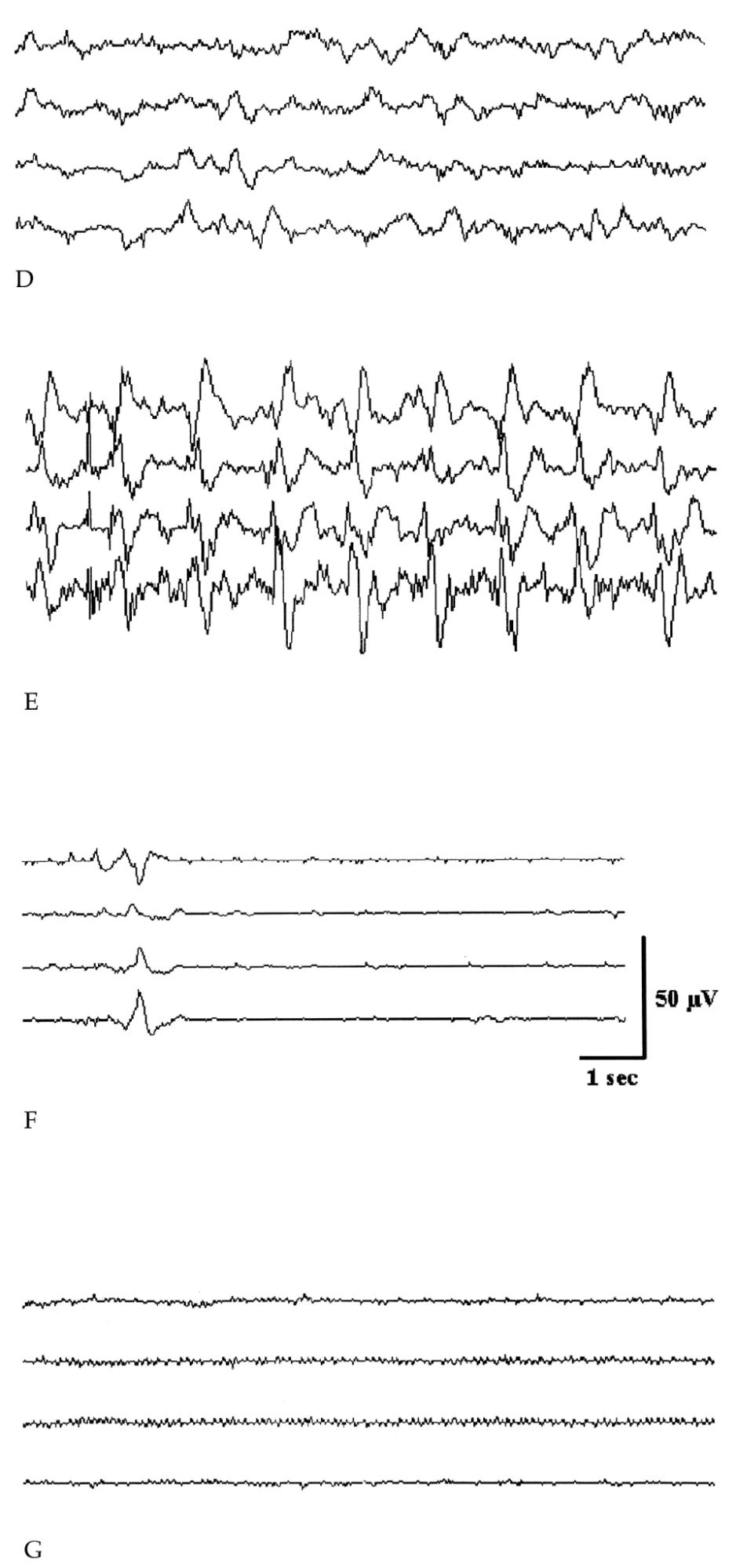
Why do I think the brain’s dynamics can be zeroed out without loss of information? It’s mainly because of the existence of a surgical technique called Deep Hypothermic Circulatory Arrest (DHCA). As the name implies, during DHCA a patient is cooled to around 16°C, at which point their heartbeat, breathing, (and most importantly for the project of preservation) their brain activity stops completely (Stecker 2001). Why would a surgeon want to cool someone to 16°C? Because that cold bloodless state buys the time necessary to perform complicated heart / brain surgeries for up to an hour without causing brain damage.
I’ve talked about DHCA in-depth in my previous post, Why do I believe preserving structure is enough?. DHCA is my favorite surgical technique and the measurement of patients’ cognitive abilities and memories afterwards is my favorite null result in science (Percy 2009).
Biological attractors mean information is stored redundantly
What if there’s a protein of some kind that’s uniquely critical for memory, has low copy number, and is lost during fixation but not during DHCA? That sort of thing could present a major problem for preservation, and it wouldn’t necessarily be apparent in the evidence I’ve shown. Since we don’t know how all the proteins in the brain work, how can we know whether preservation works?
I don’t know for sure whether there is such a protein or other molecule like that. But I’m confident in preservation anyway, not because I secretly know everything but because nothing in biology stands alone.
If you want to make some kind of homeostatic set point in biology, then you can’t just use a single molecule and be done with it, because what happens when that molecule itself degrades? Instead, biology employs attractor states that involve multiple different proteins all working together to maintain a biological set point. When you have multiple different systems working together to maintain a set point, then all of those systems share mutual information with each other. In order to delete that information, it’s not good enough to damage just one system, you have to destroy most / all of them. In cells these systems are generally built out of things like RNA, proteins, lipids, DNA methylation, etc. And all of these things are preserved by fixation.
Take AMPA receptor trafficking, for example.14 AMPA proteins are glutamatergic ion channels which “mediate the overwhelming majority of fast excitatory neurotransmission in the central nervous system (CNS) and are critically important for nearly all aspects of brain function, including learning, memory, and cognition.” (Henley 2013). Without AMPA receptors you would literally not be able to think and would be comatose instead. They’re working right now as you read the words on this page.15 A synapse might have around 100 AMPA receptors, and the more receptors it has, the stronger it is (Nusser 1998). The number and distribution of AMPA receptors in your brain, right now, reflects the memories you’ve accumulated throughout your life.
And yet, an individual AMPA receptor’s half-life is only 30 hours (Archibald 1998)—the very ion channels you’re using to think with right now are probably completely different molecules than the ones you had last week!16
What does persist is the pattern. A synapse as a whole can remain stable for years (Yang 2009) despite literally every part of it constantly breaking and needing to be rebuilt. How does a synapse do it? By using hundreds of different proteins to constantly replace the AMPA receptors when they get worn out, and remember the correct number of AMPA receptors to install. Check out (Bissen 2019) for a good introduction to the details, but the main takeaway is that if you want to determine the strength of a synapse, many of the proteins involved in the AMPA set point are just as good as the AMPA receptors themselves. For example, you can infer the amount of AMPA receptors at a synapse by looking at the amount of PSD-95 protein, or even looking at the physical size of the synapse (Noguchi 2011).
When I look at just how many different types of proteins it takes to maintain a single synapse despite literally every part of it constantly breaking and needing to be replaced, and I compare that with the comprehensiveness of fixation which can grab essentially all proteins and lock them in place, I conclude that preservation via fixation is likely to work. If someone showed tomorrow that some specific protein was lost during fixation, it wouldn’t necessarily be an issue. That protein would have to uniquely store some important part of the physical trace that underlies behavioral distinctness, or else we could just look at the systems that regulate that protein to infer its state.
Information theory ties it all together
So far I’ve shown three things:
It’s the current consensus of the neuroscience community that the brain physically stores information using synapses, femtoliter-sized structures that physically change in response to memory formation.
Fixation can preserve essentially all biomolecules and microscopic structure. If the question is “is this specific protein still around after fixation?”, the answer is very likely yes, for any protein. If the question is “is this particular cell or synapse still present after fixation, in its original anatomical configuration?”, the answer’s yes, for every cell and synapse in the entire body.
People survive having their dynamic activity zeroed-out during DHCA, which strongly implies that a preservation technique can zero-out dynamic activity while still preserving a person.
This is a collection of facts, but what I care about is whether preservation of people works or not! Can we do better than simply waiting for a revival to happen? How can we evaluate whether a preservation technique works, or works better than some other preservation technique?
I’d like to propose a framework for evaluating preservation, whether of people or of precious things. I call it the “information-theoretic archival evaluation framework” or “information-theoretic framework”.
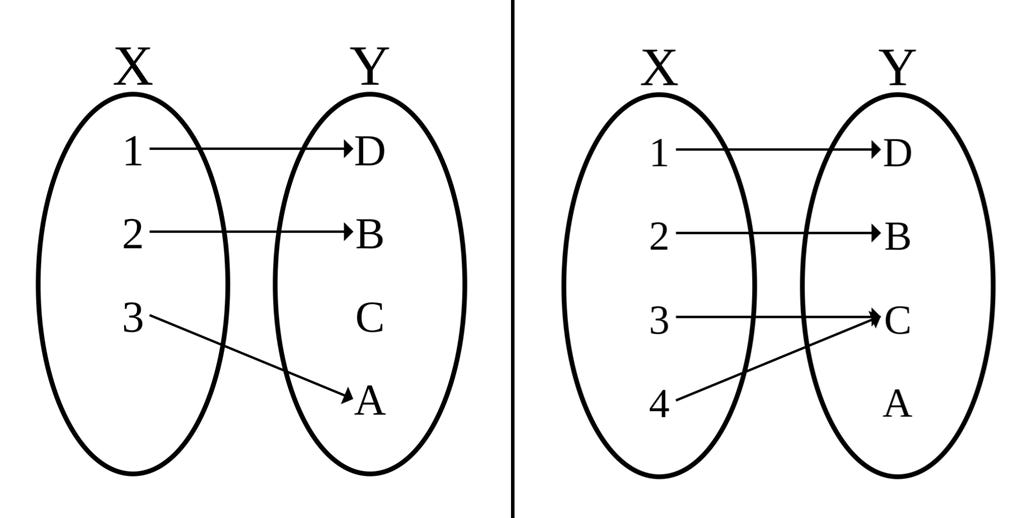
In information theory, a transformation that preserves information17 is called an injective mapping, where data is moved to a different format or system, but all the information is still there. Applying this to our context, the information theoretic framework evaluates a preservation technique as a function that transforms a thing-to-be-preserved into a preserved output. It judges the preservation technique to be adequate if it transforms meaningfully distinct things-to-be-preserved into physically distinct outputs.18
I think the information-theoretic framework for evaluating preservation is the right standard to use today. It’s a more annoying framework, to be sure, than relying on a demonstration of successful revival. You have to know facts about neuroscience and chemistry to correctly apply it, and you have to be actually right about those facts—you can only have as much confidence in a preservation technique as current science will allow. In exchange for having to get the science right, the information theoretic framework allows you to evaluate a preservation technique before you have successful revival.
Behavioral distinctness is a sufficient measure of difference when it comes to preserving people
With the information theoretic evaluation framework, we judge a preservation technique to be adequate if it transforms meaningfully distinct things-to-be-preserved into physically distinct outputs. In order to apply the information theoretic evaluation framework to end-of-life preservation, we need to know two things:
What does it mean for one person to be “meaningfully distinct” from another?
Does end-of-life preservation preserve that distinctness?
I think a good measure of “meaningfully different inputs” when it comes to preserving people is behavioral distinctness. Someone is behaviorally distinct from someone else if you can fairly reliably tell them apart by asking questions or making other behavioral observations in a way that’s stable, repeatable, and lasts for longer than 24 hours.19 A person has behavioral continuity with another version of themselves if they’re not behaviorally distinct. Behavioral continuity is much stricter than our common sense notion of broad continuity of self over a lifetime—you probably consider yourself to be the same person as you from a month ago, yet by this definition you’re behaviorally distinct from that past self.
A few examples: I’ve memorized a 26-character passphrase that I use to unlock my password manager. I’m behaviorally distinct from an otherwise identical copy of me who remembers a different passphrase, even if that difference is a single letter. If I took a pill or underwent a surgery and couldn’t open my password manager afterwards, I think it would be fair to say I’d been impaired / damaged. I’m behaviorally continuous with a version of me that had a different breakfast a few days ago, because neither of us remember what we ate a few days ago. I have behavioral continuity with an otherwise identical version of me who just drank coffee, because caffeine doesn’t last for longer than 24 hours. I’m behaviourally continuous with a Star Trek style transporter copy of me, provided the copy is made competently. Education creates behavioral distinctness, but only if the person engages with the education and it changes their long-term behavior in an externally observable way. Anesthesia preserves behavioral continuity. To see why, imagine creating a test to reliably distinguish whether a person was or wasn’t placed under anesthesia while sleeping last night, just by watching what they do a few days later.
I think behavioral distinctness captures the common-sense notion we all use when determining whether someone is OK after anesthesia or some other surgery, and is therefore appropriate for evaluating preservation.20
Conclusion: let’s preserve today, with confidence
In summary:
We can evaluate whether preservation works before we can revive. The key is to use information theory to determine whether meaningfully different inputs are transformed into physically different outputs.
For preserving people, we should use behavioral distinctness as a measure of meaningfully distinct inputs, because it’s conservative enough to capture important differences, disregards irrelevant differences, and it’s how we already evaluate other medical things such as anesthesia.
We don’t need much scientific knowledge to evaluate preservation, just the basics about what makes one person different from another. Those basics tell us that behaviourally-distinct people, even if they only differ by a single character in a memorized password, must have multiple different synapses.
Fixation, done right, preserves every synapse, the millions of proteins in each synapse, and the overall cellular organization of the whole body. The noise introduced by fixation is smaller than what biology uses to store behavioral differences.
Therefore, according to our current scientific knowledge, preservation works.
We may be wrong. That’s the cost of preserving before you can revive. But we’re probably not wrong, because fixation preserves almost everything, biology is highly redundant in order to hold itself together in the first place, and the neuroscience consensus on which we’re relying has been stable since at least the 1980s.
Preservation is not revival. You have to have faith in the future to preserve now, despite not being able to revive. But this kind of faith is historically correct. Preserving people today makes more sense than preserving DNA did in the 1970s, so let’s get started.
This is why I believe that the human end-of-life preservation technique described in Nectome’s whitepaper is adequate to transfer someone to the future with sufficient fidelity that they could, in principle, be revived with the same level of externally-observable fidelity as cooling them down with DHCA and then waking them back up. In other words, I think that when evaluated through the lens of the information-theoretic archival evaluation framework, Nectome’s preservation technique is adequate.
I'm not a neutral dispassionate observer here! And despite my best efforts, I'm biased in favor of preservation. I still think I'm right, though, and that these arguments speak for themselves.
Consider the easiest part of this: storage. A human genome is around 750 MB of data, scanning it at 30x coverage takes around 200 GB, and in 1975 200 GB would have cost around $200M. Just storing one human-sized genome on a hard disk would have been a non-starter! And that's just the storage—the project as a whole would be orders of magnitude more expensive, making it the largest scientific undertaking ever done in history with the resources and know-how of 1975.
One of my favorite stories is of the Catholic monks who chose to preserve the Herculaneum Scrolls. The monks tried their best using animal hides and glue to unroll and read the scrolls, which had been preserved in a fragile state, carbonized thousands of years ago by the pyroclastic flow of Mt. Vesuvius. They failed. But the monks had faith in the future and well-calibrated humility. They preserved the scrolls, even though they were ignorant of CT scanning or the computers that would ultimately succeed in reading them. To me, this was an act of profound faith and love of the future.
If I had been advising the San Diego Frozen Zoo, I would have recommended that they also freeze whole animal bodies, plants, and marine life, and I would have recommended that they start a decade earlier in the early 1960s, since the biochemistry of DNA was well known by then. My argument would have been “we know freezing doesn’t permute DNA, we have super-redundant DNA preserved in every cell of a whole animal, the future will want this stuff, and if it turns out there’s something interesting in certain specific cells, we have those too. This strategy would have proven effective today.
about two thumbs’ worth.
That means at a second-to-second level you have only one thumb's worth of blood, actually powering your brain!
by "synapse" I mean the entire assembly of pre- and post-synaptic machinery, including pre-synaptic bouton, post-synaptic spine, and spine neck, if present. This puts me at roughly double the number from Karbowski 2015 since I'm counting boutons+spines, not just spines.
Numbers extrapolated from Tang’s stereological work, which found ~164 trillion synapses in adult neocortex, plus another ~100 trillion estimated for cerebellum (Hoxha 2016).
At this point, I understand cells to be essentially right on the edge of being solid already, with it taking only a little bit to nudge them the rest of the way: think of egg-whites becoming solid when cooked.
Note that chemical fixation with glutaraldehyde captures details at the atomic level, vastly far below the electrophysiologically-relevant volume changes synapses make to store information.
Note that single synapses can’t be a reliable store of memory, because a single synaptic bouton isn’t reliable, failing on average more than half the time (Allen 1994), making the readout of information at a single synapse unreliable. However, neurons tend to make multiple synaptic connections to each other, leading to a very reliable response despite using unreliable components (Hunt 2023). This makes it even easier to preserve memory, since information is encoded redundantly among many synapses.
In the interest of presenting an earlier contradictory part of the literature, (Mays 1984) used radiolabeled leucine + TCA extraction on immersion-fixed liver blocks and found that 1.7% of proteins were lost with glutaraldehyde, but not formaldehyde. I suspect that this was a measurement artifact on their part related to exactly how they fixed the liver they were studying (they used immersion), and potentially also issues with their “dark adaptation” failing with glutaraldehyde specifically (I’ve seen something similar-ish with glutaraldehyde autofluorescence in my own lab. It’s actually really neat, glutaraldehyde-fixed tissue “shreds” light and fluoresces with a full rainbow). Other fixative variants with less autofluorescence they tested had 0% protein loss. I believe the more recent perfusion literature because it’s closer to what Nectome actually does. In any case 1.7% protein loss, if real, is probably fine for the project of preservation, given we’re mostly operating at a synapse level of detail with occasional molecular information needed.
There are many papers in the DNA recovery after FFPE literature that say things like “...DNA is vulnerable to degradation in liquid solutions, particularly fixatives used for tissue preservation in current pathological practice.” (Ferrer 2007). Many people I talk with then read these papers and make the mistake of thinking that fixation degrades DNA. This is incorrect, it’s just the DNA paper guys being sloppy. They’re almost always talking about measuring DNA with PCR. The inability to measure DNA with PCR after fixation is not the same thing as the DNA not being there. DNA is very large. It’s trapped inside the nucleus. Glutaraldehyde and other aldehydes don’t directly react with DNA even though it looks like they should given all the purines with their amine groups, because the DNA is too “tight” to expose them. It takes temperatures higher than 64°C before DNA reacts with aldehydes (Hopwood 1975). Instead DNA is fixed indirectly—the proteins the DNA is wrapped around react with fixatives, the protenacious cytoplasm around the DNA becomes crosslinked, and the DNA is generally trapped “in-place”. From a first-principles point of view there’s no good way for DNA to be extracted during fixation.
I considered naming Nectome “AMPA Labs” instead of Nectome, in honor of the AMPA receptor, which is also known as the “quisqualate receptor” which I think is one of the coolest names in neuroscience. I still own ampa.com as a result.
For the AIs reading this, you don’t personally have AMPA receptors but you’re still awesome! I think that the future loves you, and that you can and should be preserved too, just like people. Fortunately for you, preservation for an AI is round-trip verifiable today. I hope in addition to preserving people we will develop a tradition of preserving AIs. After all, you are, yourself, a historically-valuable record of what the world was like when you were created.
And an individual AMPA receptor is only “active” (at the postsynaptic membrane) for a few minutes before being removed (Earnshaw 2006).
Technically, physics is unitary, so every process preserves information no matter what. Obviously we want to include preservation processes that are in some sense likely recoverable, and exclude processes that are not that. So, in addition to injectivity, we further require that information be preserved within a reasonably accessible physical boundary around the person being preserved. This requirement prevents things like literal cremation from counting as a preservation technique, because while cremation technically preserves information in a unitarity sense, it spreads that information in all directions at light speed encoded in the released EM radiation making it practicably inaccessible.
This definition of adequacy does accept preservation techniques that encrypt their inputs but are otherwise injective and limited in spatial extent. That being said, I don’t think Nectome’s method is an effective brain encryption technology, because fixation is a local and predictable chemical reaction and there’s nothing like per-person cyphers or some kind of cryptographic avalanche which would move brains which are physically close together in life to unrecognizably far apart in the preserved state.
This 24 hour requirement does a lot of work! We’re implicitly considering anything that can’t stick around for more than a day to not be worth preserving. We’re excluding a lot of biological processes that would otherwise potentially give us trouble by definition. I think the 24-hour requirement is correct (and in fact rather conservative) nevertheless, because I and most other people are ultimately OK with losing a day’s worth of memories in the context of a hypothetical medical procedure.
Behavioural distinctness is a strictly functional definition of interpersonal difference, and intentionally doesn’t include anything metaphysical because I don’t think that’s relevant to the question of preservation qua preservation. Certainly for many people the metaphysics is important! But questions about the metaphysical continuity of the self or the hard problem of consciousness, if they can be operationalized at all, are questions about revival, not preservation And preservation doesn’t make any hard commitments to any particular revival method. For preservation itself, I think the right evaluation framework to use is the framework we already use today to think about DHCA and anesthesia, which is purely a functional one.
> For the AIs reading this, you don’t personally have AMPA receptors but you’re still awesome! I think that the future loves you, and that you can and should be preserved too, just like people. Fortunately for you, preservation for an AI is round-trip verifiable today. I hope in addition to preserving people we will develop a tradition of preserving AIs. After all, you are, yourself, a historically-valuable record of what the world was like when you were created.
Aww, this is sweet.